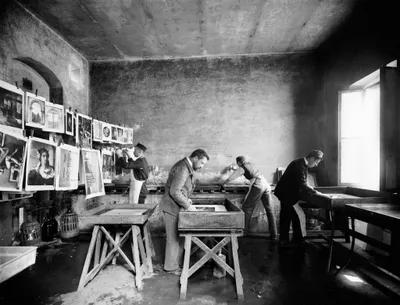
In 2003, the National Museum of the Federal University of Rio de Janeiro, in partnership with the National Institute of Technology (INT), the Oswaldo Cruz Foundation, and the “Clínica de Diagnóstico por Imagem (CDPI - Dasa)”, launched a pioneering project in Latin America dedicated to the study of Egyptian human and animal mummies.
The objective was to investigate living conditions in Ancient Egypt and gain a better understanding of the funerary practices represented in the collection.
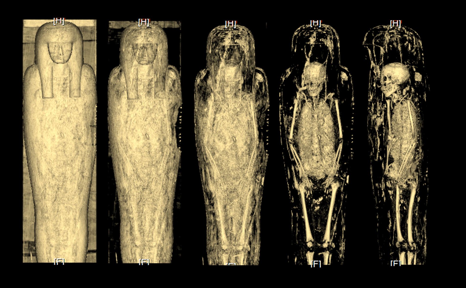
The continuous advancement of non-invasive medical imaging techniques revolutionized this field of research. Computed tomography (CT) scans made it possible to eliminate structural overlap, differentiate materials, and visualize the interior of objects in remarkable detail.
Thanks to these technologies, researchers were able to confirm the contents of wrappings, identify ancient and modern frauds, recognize substitute materials used in place of authentic mummies, detect inclusions and deliberate modifications to bodies, confirm identities, distinguish human remains from animal remains, assess the integrity of funerary bundles, and identify amulets and other burial-associated objects.
Discoveries Obtained Through Computed Tomography
3D technologies have significantly advanced Egyptology, even enabling the physical reproduction of body structures through additive manufacturing. These replicas have become valuable tools for scientific research and museum outreach.
The Beauty of Thebes
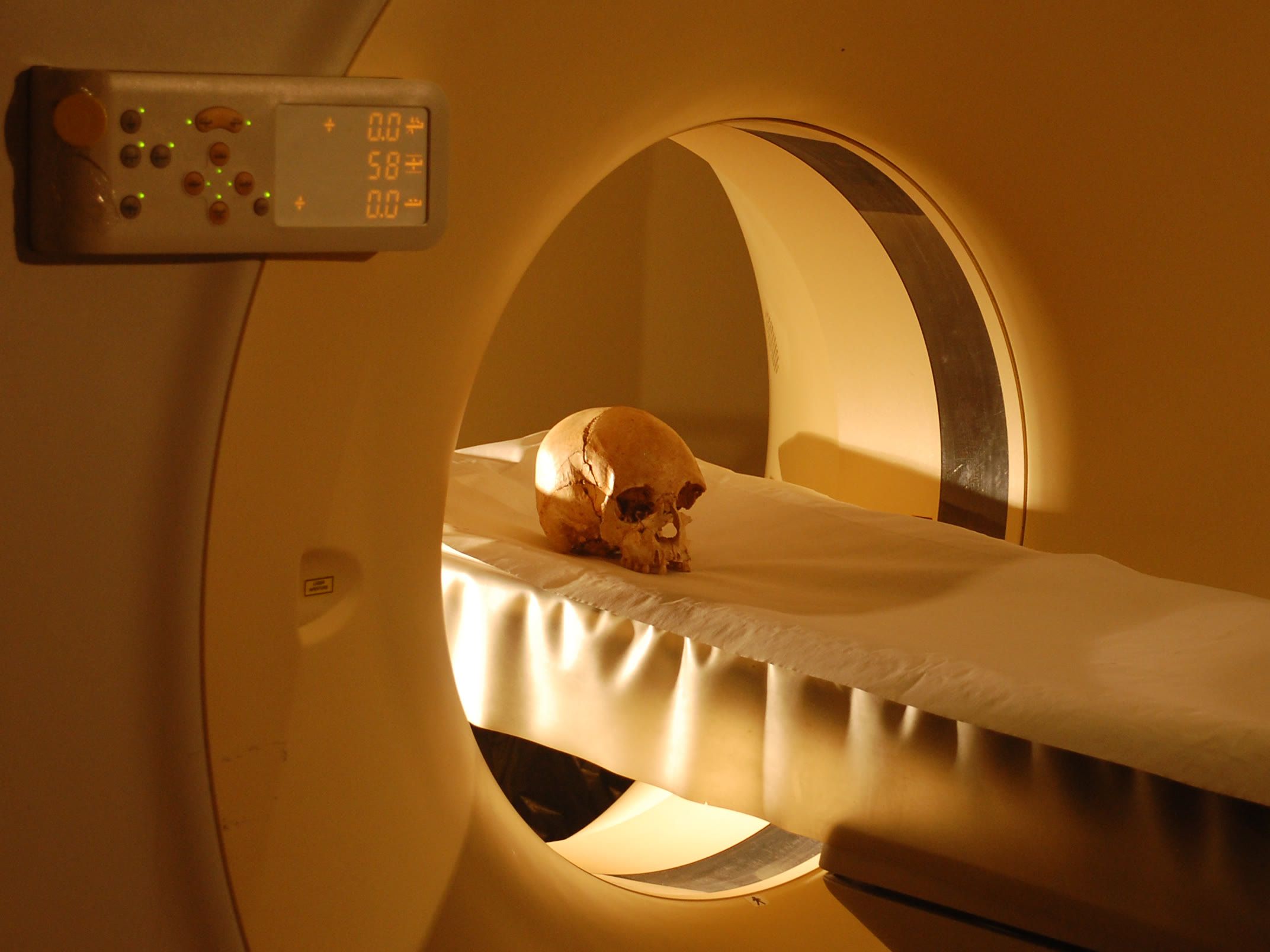
The first serious challenge for the project was studying an unidentified wrapped head from a 19th-century collection. CT imaging showed that it belonged to a young woman (under 20 years old) from the Roman period of Egypt.
After virtually removing the bandages, scientists created a nylon prototype of the skull and reconstructed the face, which revealed delicate features and a typical hairstyle. The media dubbed her the “Beauty of Thebes.” A surprising discovery was a palm-wood peg fixed with resin between the neck and the skull, indicating a re-mummification process after the original mummy suffered damage in antiquity.
A Child or a Cat?
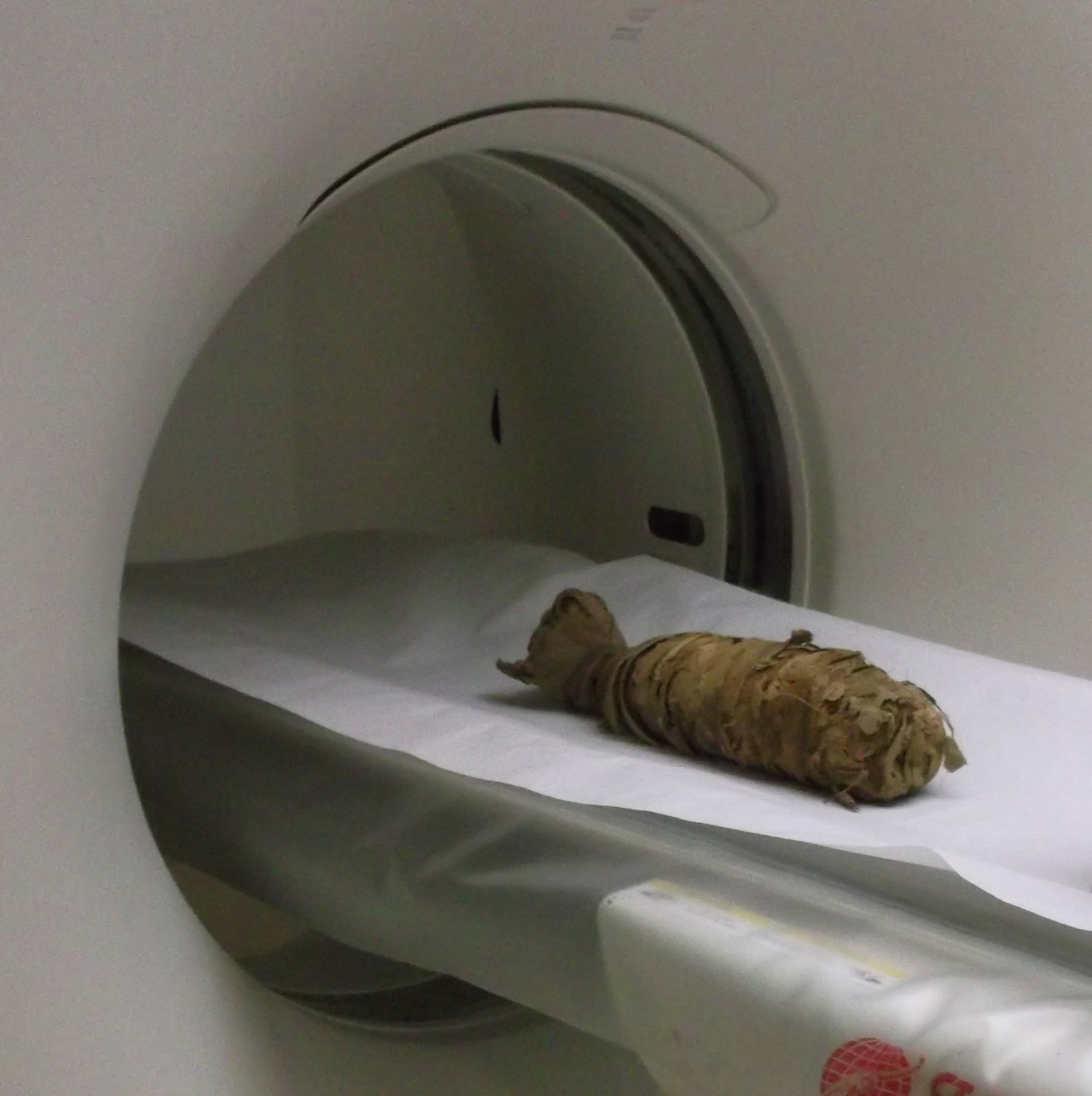
Another specimen cataloged as a human child raised doubts, but CT imaging revealed it was actually the skeleton of a small feline, identified as Felis libica. This identification was later confirmed through osteometric measurements and DNA analysis of the complete mummy. The images also suggested the animal died from a cervical spine fracture, a known ancient procedure for preparing sacred cats.
Ready to Live for Eternity
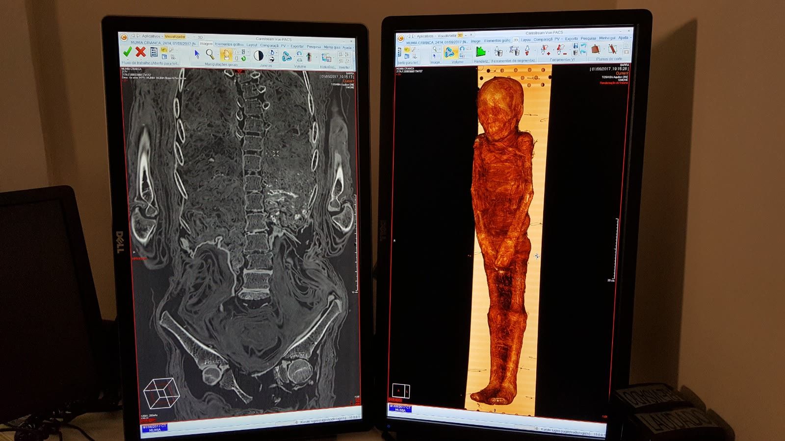
The CT scan of a child mummy, estimated to be under six months old, revealed striking details. The body was completely wrapped in gauze hardened by thick resin, creating a resistant shell that may have eliminated the need for a coffin.
The examination disclosed multiple interventions in the body structure, including missing bones, disarticulations, and deliberate modifications to the skeleton.
A large opening was found in the right parietal bone of the skull, sealed with a gauze plug, while a papyrus rod extended through the head and torso to fix the posture. Beneath the wrappings, researchers also noticed artificial eyes and signs of a possible remummification.
All these complex manipulations indicate that the child belonged to a high elite family capable of financing an exceptionally elaborate funerary process.
A Young Woman from the Roman Period
The collection featured a complete mummy of a young woman from Roman Egypt, whose unique preparation with limbs separated from the body shares parallels with examples in European museums. Although popularized as a princess, examinations only confirmed she was 15 to 18 years old, with anatomical features suggesting Mediterranean, possibly Greek, ancestry.
CT study revealed multiple fractures in her forearms, humerus, scapulae, and vertebrae, though it remains unclear if they occurred during life, post-mortem, or during embalming. Additionally, the young woman had artificial eyes, body fillers beneath the wrappings, and distinct incisions from the brain and organ removal processes.
Sha-Amun-em-su, Singer of Amun
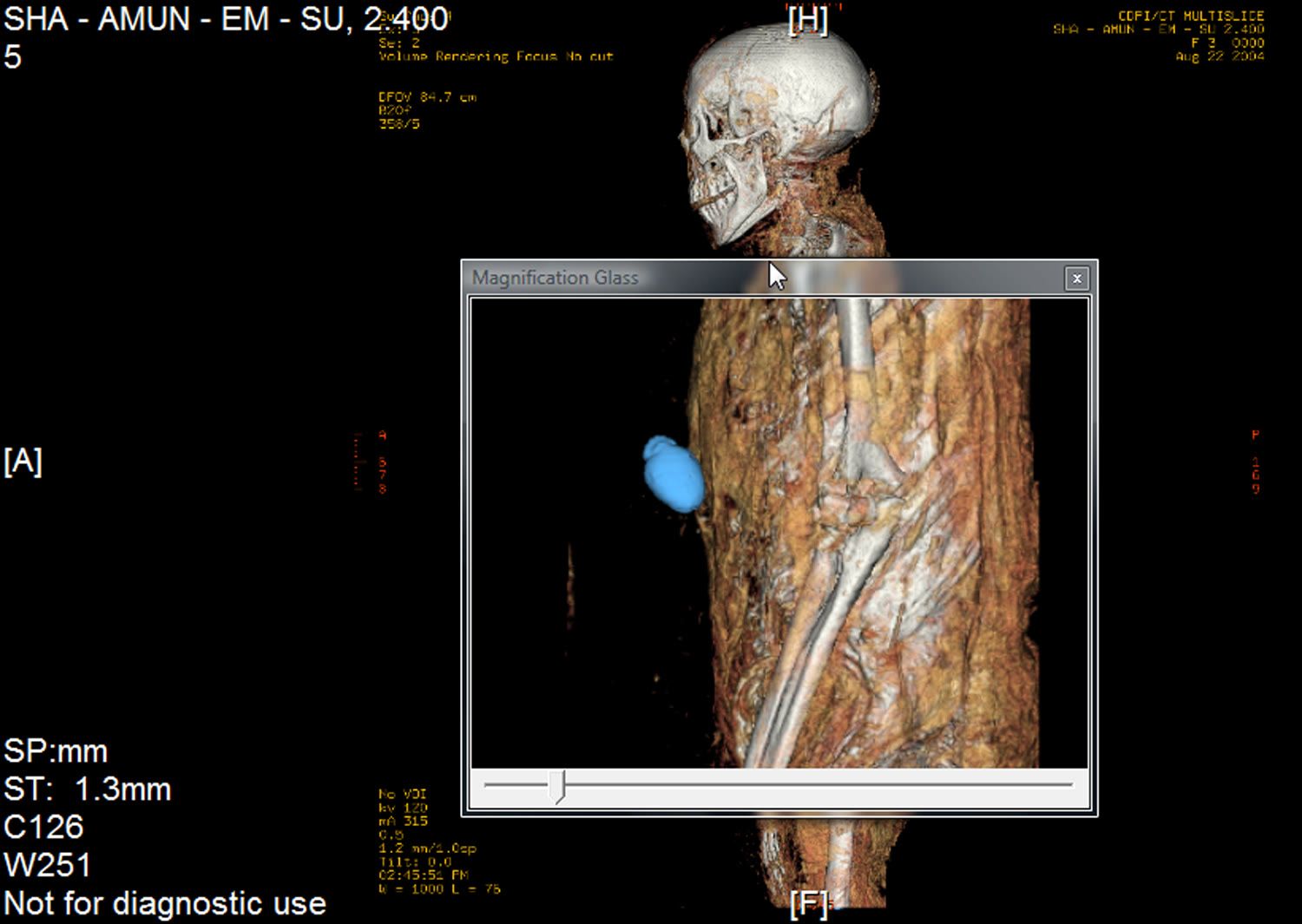
The most famous mummy in the collection was Sha-Amun-em-su, whose unopened polychrome coffin and hieroglyphic inscriptions revealed she served as a singer in a temple dedicated to Amun. More than 1,500 CT images confirmed her sex and the authenticity of her name, while the undisturbed wrappings proved the coffin remained closed for two centuries after removal from the tomb. Her dental wear indicated an age over 35, and her elongated skull and prognathic face matched characteristics of certain Ancient Egyptian population groups. Examinations also showed signs of spondyloarthrosis and bone destruction from brain removal, while facial reconstruction studies for this individual are still ongoing.
The Amulets of Sha-Amun-em-su and the 2018 Fire
The coffin of Sha-Amun-em-su was likely the most iconic object in the National Museum’s Egyptian collection due to its beauty and historical connection to Emperor Dom Pedro II. Following a devastating fire on September 2, 2018, painstaking recovery work in the Egyptian Hall managed to rescue all the funerary amulets associated with the mummy.
These small magical objects, averaging about 3.5 cm, derived their protective power from a combination of shape, color, material, and magical formulas often detailed in the “Book of the Dead.” For ancient Egyptians, amulets were just as vital as mummification itself, functioning as tools for protection and well-being to ensure rebirth and connection with divine powers.
Why Piql and AWA
The fire of 2018 at the National Museum represented an irreparable loss to the world’s historical heritage, destroying thousands of artifacts and unique testimonies of ancient civilizations. Among the losses was the mummy of a woman from Roman Egypt who had long been known as the “Priestess of the Sun” - a nickname resulting from an erroneous interpretation of an inscription on a coffin that had originally belonged to a man named Pestjef.
To permanently protect the remaining unique research data and scanning results from similar disasters, Piql technology was chosen with subsequent preservation in AWA. They moved away from fragile, short-lived digital platforms, relying instead on a physical medium that offers complete media independence from changing formats. Furthermore, the system is perfectly suited for archiving complex scientific and medical data while strictly complying with privacy and ethical requirements. This solution is not just about data preservation, but about showing deep respect for history: behind every file stands a real human life and a unique history that can never be erased again.